Examinations during pregnancy
PRENASCAN
PRENASCAN is a non-invasive prenatal test of chromosomal defects in the fetus. The examination mostly focuses on trisomy of chromosome no. 21 - Down syndrome, no. 18 - Edwards syndrome and no. 13 - Patau syndrome. The test can in addition determine the risk of other chromosomal defects including defects of sex chromosomes.
When is PRENASCAN recommended?
PRENASCAN is recommended after pregnancy week 10 after genetic consultation, for example in the following cases:
- increased risk of trisomy 21, 13 and 18 is detected in biochemical screening
- atypical screening result with borderline risk
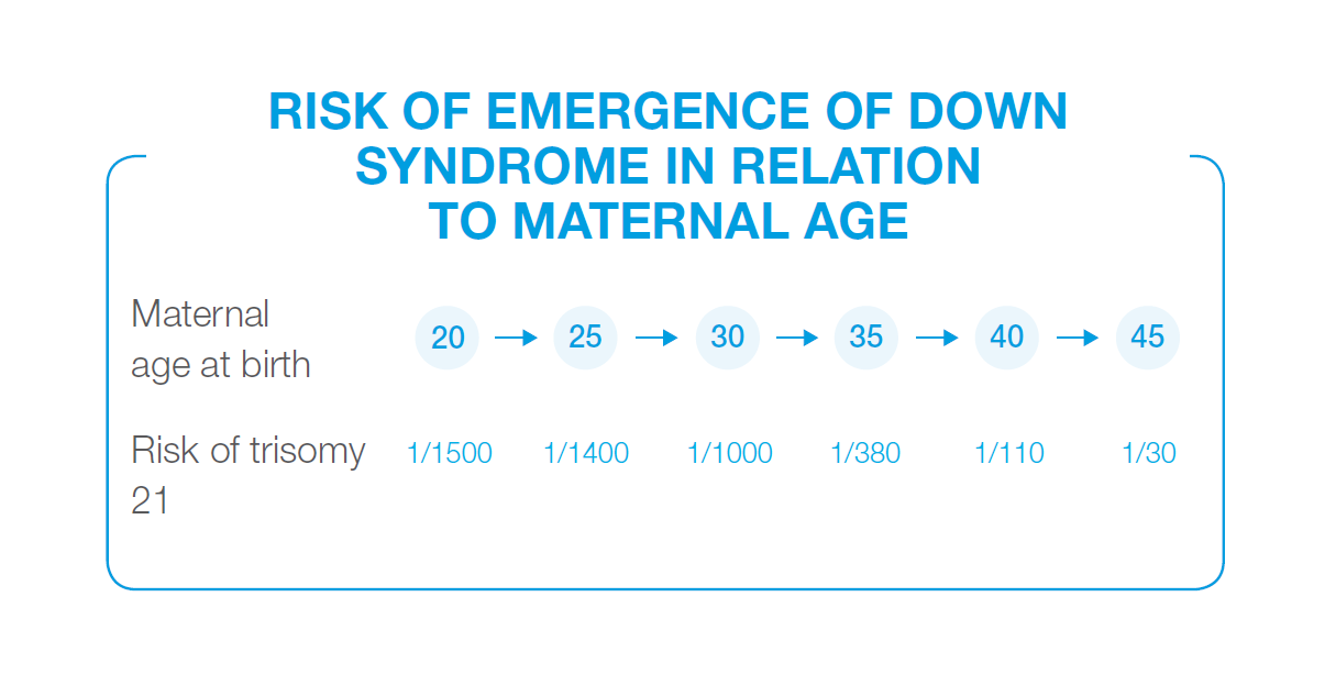
- age of the mother over 35 years
- assisted reproduction
- fears about the outcome of pregnancy
- suspicion based on examination of family tree
PRENASCAN is not recommended as the test of first choice for variations in forming fetal organs that are detected in ultrasound examination. In such cases there is a reason to examine fetal tissue directly that is collected with:
- chorionic villus sampling (CVS)
- amniocentesis (AMC)
- examination of the fetus with methods of molecular genetics (microarray)
What are chromosomes and in what cases do fetal chromosomal defects emerge?
- chromosomes are blocks of genes of typical shape and number that are stored in nuclei of all cells
- humans have 23 pairs of chromosomes = 46 chromosomes
- one member of the pair comes from the mother and the other from the father
- mature sex cells - egg and sperm cells - have half of the number of chromosomes, which are paired at conception
Chromosomal defects and their consequences:
Trisomy = three copies
- occurs when a “trio” develops at conception instead of a pair of chromosomes
- means that the person in question has 47 chromosomes in each cell of each organ and not only 46
- mostly develops randomly at conception
- occurs most frequently in chromosomes number
- 21 - Down syndrome
- 18 - Edwards syndrome
- 13 - Patau syndrome
- the risk increases with the age of the mother
Structural defects
- only affect parts of chromosomes
- with loss (deletion) of genetic information
- with excess (duplication) of genetic information
- they can be the cause of serious illnesses that affect multiple organs
There is no efficient prevention or specific treatment of chromosomal defects and life expectancy of the affected patients is often poor.
What is the course of procedure with PRENASCAN?
PRENASCAN is based on examination of cell-free fetal DNA that can be found in the blood of the mother. Blood is collected from the patient and sent to the lab. The results of the test are available in 2 to 4 weeks.
Methodology:
- deoxyribonucleic acid (DNA) carries genetic information and is stored in chromosomes in each cell nucleus
- blood consists of cells and plasma
- freely circulating fragments of DNA can be detected in plasma, they are released from decomposed cell nuclei
- mixture of DNA fragments of mother and fetus can be found in the plasma of pregnant women
- each one of the millions of DNA fragments in maternal plasma can be paired with the chromosome from which it originated with the method of massive parallel sequencing (MPS), which can in addition detect excess of DNA of a particular chromosome that is caused by fetal trisomy
What is the test efficiency compared to invasive fetal examination?
- MPS that is used in PRENASCAN detects trisomy 21 and 18 with accuracy that is close to direct examination of amniotic fluid cells or placental cells.
- Diagnostic accuracy for rare trisomy 13 is almost 99 %.
- Gender of the fetus is determined with evidence of DNA chromosome Y.
- Efficiency of PRENASCAN for twins is similar as for singleton pregnancy.
Why do physicians in some cases recommend invasive examination with risk of complications in the pregnancy when the reliability of PRENESCAN is this high?
PRENASCAN is not a universal prenatal test. There are still reasons in some cases for:
- collection of amniotic fluid
- chorionic villus sampling
- direct examination of fetal cells with methods of molecular genetics
Diagnostic sensitivity of more than 99 % cases of most common trisomy:
- is comparable with direct invasive examination of fetal tissue
- false negativity of the test is not completely excluded though
Specificity of the test is about 99%
- there is a risk of false positive result of this test
- if there is a positive result, it is recommended to verify it with invasive examination
- of chorionic villi (CVS)
- of amniotic fluid (AMC)
Results of the test can be distorted when:
- mother has a chromosomal aberration herself
- test was carried out in early pregnancy (when there is still not enough of cell-free DNA in maternal blood)
- quality of cell-free DNA prevents the outcome of the test
- fetus has a rare combination of chromosomal aberrations (i.e. chimerism, microduplication and microdeletion)
- foreign DNA is present in maternal blood (i.e. after transfusion or transplantation of stem cells)
